


Most Amazing Pictures of 2019’s Nikon Photomicrography Competition.
Nikon’s annual Small World photo competition highlights the beautiful elements of nature that can only be seen through the lens of a microscope.
Now in its 45th year, the competition drew more than 2,000 entries from scientists in over a hundred countries, which were narrowed to the 20 winners announced today.
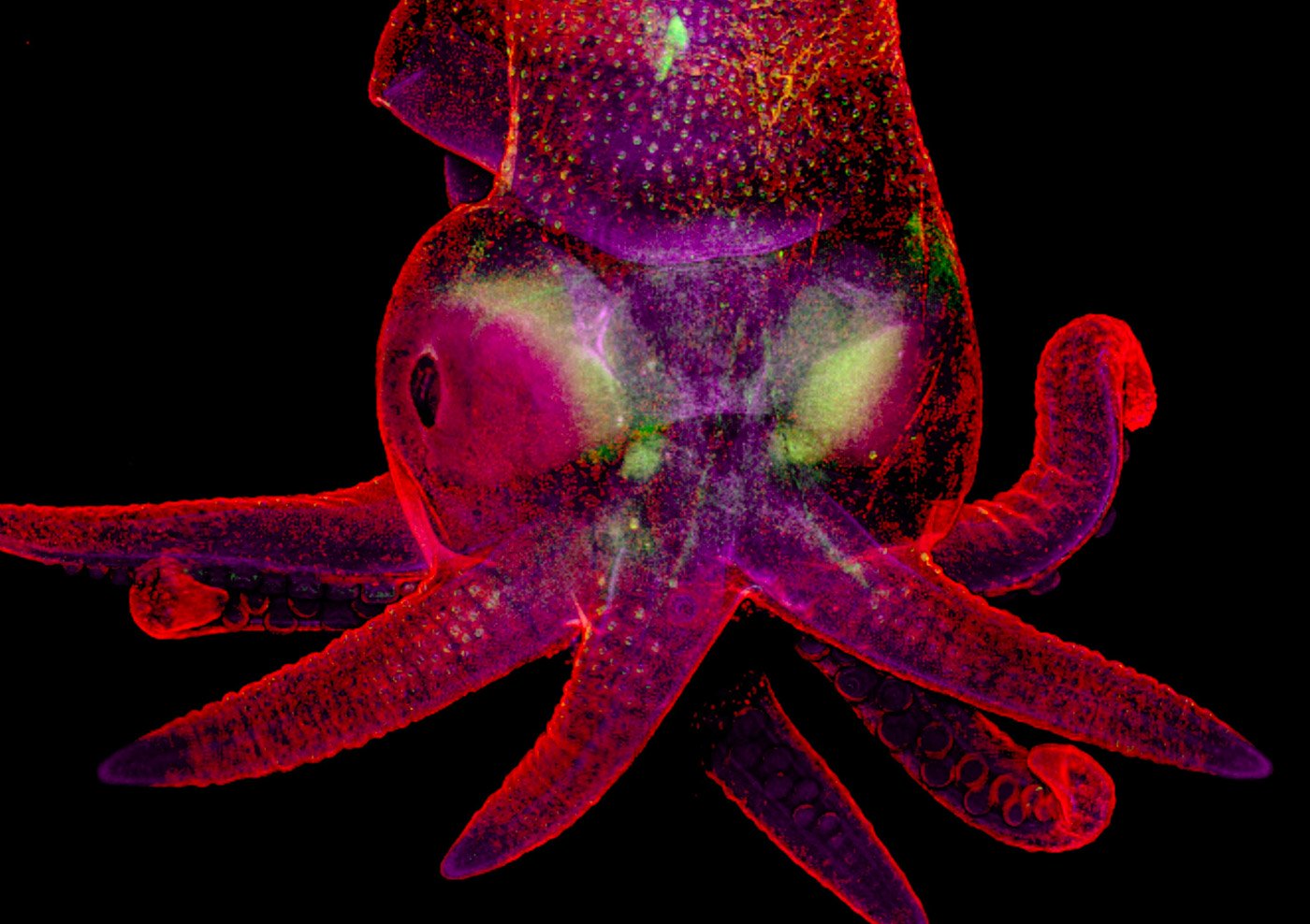
The first prize went to this image of a turtle embryo, shot by microscopy technician Teresa Zgoda and recent university graduate Teresa Kugler, using a combination of fluorescence and stereomicroscopy. The striking capture beautifully combines their passion for art and science.
They captured the image using fluorescence and stereo microscopy and image-stitching.
Kugler said: “Microscopy lets us zoom in on the smallest organisms and building blocks that comprise our world – giving us a profound appreciation for the small things in life that far too often go unnoticed.”
Zgoda added: “We are inspired by the beautiful images we see through the microscope.
“It’s humbling and deeply fulfilling to be able to share that science with other people.”
H/T: Fast Company


Life on Earth is a miracle and it all started so small.
It`s just WONDERFUL, what the fotos show!!
Juergen from Loy (PJP)
LikeLiked by 1 person
Amazing! 😀
LikeLiked by 1 person
Reblogged this on UN ESPRIT SAIN DANS UN CORSAGE and commented:
ET S’IL VOUS PLAÎT, NE MANGEZ PAS LE PETIT COCHON À DROITE !
LikeLiked by 1 person
Superbes images !
LikeLiked by 1 person
A world so fascinating ! Incredible!
LikeLiked by 1 person
Wow!
LikeLiked by 1 person
Amazing. This is such an interesting blog. I’m glad I found you.
LikeLiked by 1 person
A reblogué ceci sur Perfect-Style-Knower.
LikeLike
Amazing pictures
LikeLiked by 1 person
I could look at these everyday
LikeLiked by 1 person
totally awesome!
LikeLiked by 1 person
These are truly amazing. Thank you for sharing.
LikeLike